By The Pawline veterinary teamVeterinary-reviewed by Alastair Greenway, BVM&S MRCVS · 23 August 2026
Electrophoresis separates blood proteins by how fast they travel through a gel, producing a trace rather than a number. It is ordered when [globulin](/lab/globulin) is raised and the question is WHY — and the answer is in the shape of the trace: a narrow spike and a broad hump mean very different things.
A standard chemistry panel measures total protein and albumin, and works out globulin by subtracting one from the other. That is useful, but blunt: "globulin" is not a substance. It is everything in the blood that is not albumin — hundreds of different proteins, including every antibody the animal makes.
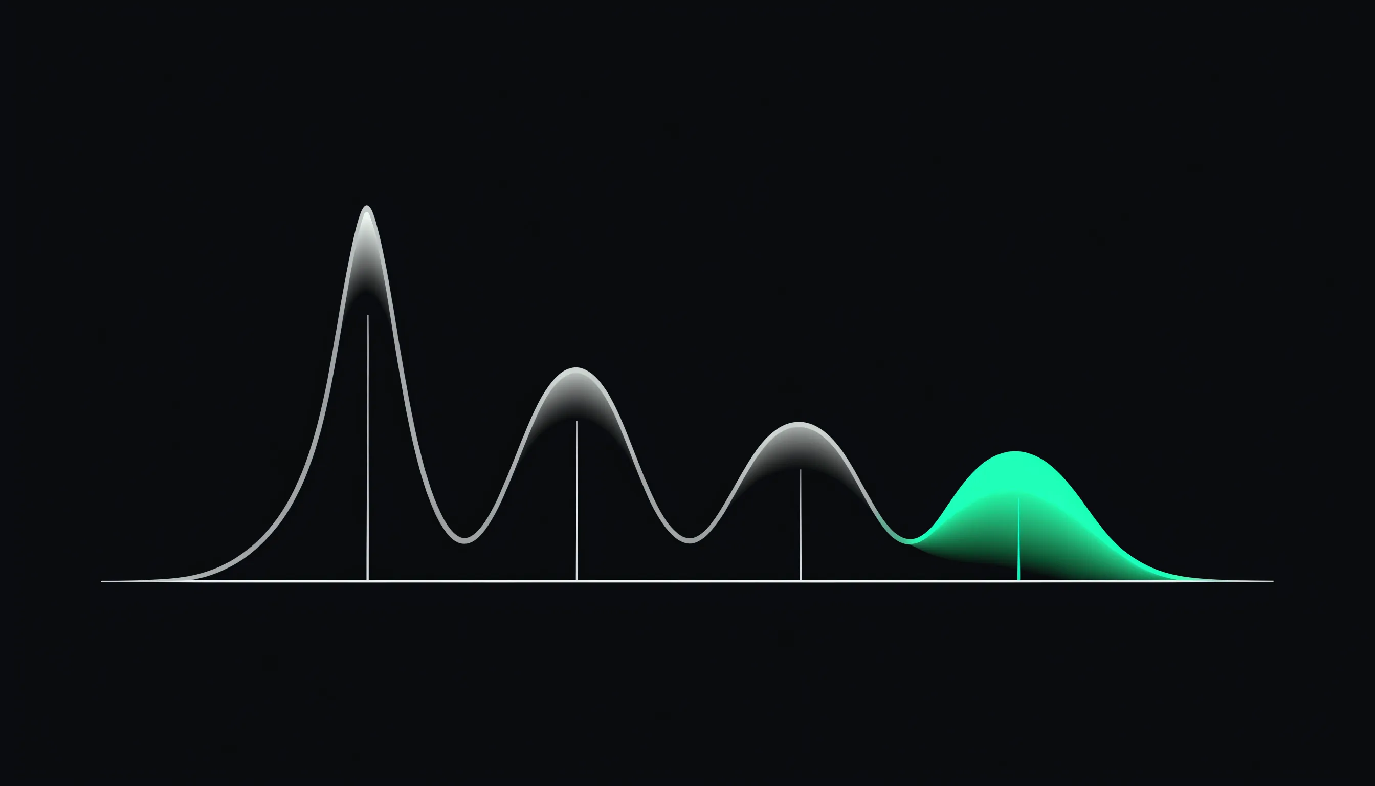
Electrophoresis separates serum proteins into fractions; the shape of the gamma region is the finding.
Electrophoresis unpacks it. The serum is drawn through a gel by an electric current, and proteins separate by how quickly they travel. The result is a trace with peaks rather than a number, conventionally divided into albumin and then the globulin fractions — alpha-1, alpha-2, beta and gamma.
The finding is the shape, not the height
This is the point of the test, and it is why a raised globulin on its own is not enough to act on. Two animals can have an identical globulin and traces that mean entirely different things.
A broad, rounded rise in the gamma region — a polyclonal gammopathy. Many different plasma cells are making many different antibodies at once. This is the immune system doing what it is supposed to do, on a large scale, and it points at chronic stimulation: long-running infection, persistent inflammation, some immune-mediated diseases. It says the body is responding to something; it does not say to what.
A narrow, tall spike — a monoclonal gammopathy. One clone of plasma cells is making one antibody in enormous quantity, which is a fundamentally different biological event. It usually migrates in the gamma or beta region, and it raises the possibility of a myeloma-related disease: multiple myeloma, a plasmacytoma, or certain immunoglobulin-secreting lymphomas and leukaemias. IgG and IgA spikes are the commonest in dogs and cats; IgM is rare.
A spike on electrophoresis is a strong pointer, not a diagnosis. Immunofixation — a further step on the same sample — is what confirms a spike is genuinely monoclonal and identifies which antibody class it is.
The other fractions
The gamma region gets the attention, but the earlier peaks carry information too. A rise in alpha-2 reflects the acute-phase response — proteins the liver makes rapidly during inflammation — which is a different timescale from the antibody response and can be read alongside CRP in dogs.
A low albumin peak with raised globulins is the pattern behind a low albumin:globulin ratio, and the trace shows which globulins are responsible.
Cat vs dog
The method and the interpretation are the same, but there is one feline context worth knowing.
In cats, feline infectious peritonitis classically produces a raised alpha-2 fraction together with a polyclonal gammopathy — the acute-phase response and the antibody response at once. That pattern is typical but not specific: other inflammatory diseases produce it too, and electrophoresis cannot diagnose FIP by itself.
There is a further wrinkle worth stating, because textbook accounts can imply a cleaner rule than exists: monoclonal spikes have been reported in cats with FIP. A narrow peak in a cat is therefore not automatically a neoplastic finding, and the trace is interpreted with everything else rather than read as a verdict.
About the reference range
This page shows no range strip, and the reason is more fundamental than usual. Electrophoresis does not produce a single value to place on a scale — it produces a trace, reported as the sizes of several fractions plus a pathologist's description of their shape. Methods differ too (agarose gel is the common one; capillary zone electrophoresis is faster and used by some laboratories), and fraction values are not directly comparable between them.
Read the laboratory's own report and, in particular, its comment: on this test the interpretation is the product.
What to do next
Electrophoresis is a second-line test, ordered to explain a raised globulin or a low albumin:globulin ratio that has already been found. If it has been recommended, it is worth asking your vet whether immunofixation will be run should a spike appear — it is the step that turns "there is a narrow peak" into a characterised finding, and running it on the same sample avoids a second blood draw.
This page is educational and is not a diagnosis. Lab results are interpreted in the full clinical context by your veterinary surgeon; a single value rarely tells the whole story.